
This article is an excerpt from the Shortform book guide to "The Body Keeps The Score" by Bessel van der Kolk. Shortform has the world's best summaries and analyses of books you should be reading.
Like this article? Sign up for a free trial here .
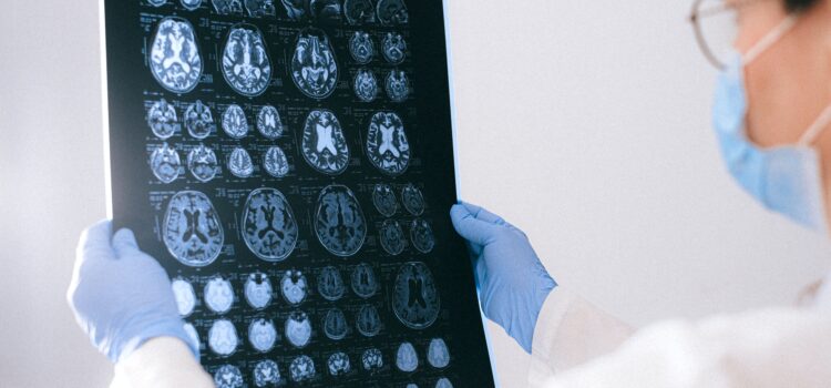
How does PTSD brain imaging work? Can we see how the brain reacts to trauma using imaging?
PTSD brain imaging allowed scientists to look at the physical way that the brain processes and reacts to trauma, including how PTSD works. This enables scientists to make new discoveries in the way the brain works, including how we handle trauma.
Read more about PTSD brain imaging and related discoveries.
PTSD Brain Imaging Reveals Trauma’s Effect on the Brain
The advent of brain-imaging technology in the early 1990s gave scientists new insight into the way brains process information, memories, sensations, and emotions. Previous research measuring brain chemicals (like serotonin) revealed what fueled brain activity — like trying to understand how a car engine works by studying the gasoline — whereas this new technology was like looking inside the engine.
With these tools, researchers learned that trauma leaves an imprint on the mind, brain, and body that has long-term effects for how you survive in the present. In the ways that trauma impacts perceptions, imagination, relationships with other people, and other mental processes, it not only changes how and what you think, but your capacity to think.
As a result, traditional talk therapy is typically insufficient treatment for trauma, because it doesn’t repair the physical and hormonal responses that cause trauma survivors to be hypervigilant to threats. Instead, in order to heal the survivor needs to internalize — mentally and physically — the understanding that the trauma is in the past, so that she can live in the present without being haunted by the event. We’ll explore ways to achieve this in later chapters.
Parts of the Brain Affected by Trauma
The author enlisted eight trauma survivors to participate in a study in which he and other researchers would recreate scenes of their trauma — essentially triggering a flashback — as they had their brains scanned to see the reactions.
The researchers found that participants’ amygdalas (the cluster of brain cells that distinguishes threats) activated in response to images, sounds, or thoughts related to their trauma as if the danger was present — no matter how many years had passed since the traumatic event actually occurred. The amygdala rings the alarm of impending danger, and the body releases stress hormones and nerve impulses that make blood pressure, heart rate, and oxygen intake rise in preparation for fight or flight. PTSD brain imaging is able to show these changes.
Flashbacks Inhibit Speech
PTSD brain imaging shows areas where the brain is inhibited. During the brain scan experiment, the researchers also found that a part of the brain called Broca’s area was debilitated during a flashback.
Broca’s area is a speech center in the brain that is key to being able to articulate your thoughts and feelings; Broca’s area is often affected in stroke victims. The experiment showed that trauma flashbacks can cause temporary physical effects — the brain’s speech control center going offline — that are comparable to strokes.
But trauma survivors’ difficulty with speech lasts beyond the confines of a flashback; years after a traumatic event, survivors struggle to express what happened. With time, most develop what the author calls a “cover story,” an outline of the basic events, but these cover stories fail to capture the depth of the experience.
Flashbacks Trigger Intense Emotions While Debilitating Logic
While speech is inhibited during flashbacks, another part of the brain called Brodmann’s area 19 was activated in participants’ brains during the flashbacks. Brodmann’s area 19 is responsible for managing images as they enter the brain, then sending them to other areas of the brain to interpret the images’ meanings. This is another interesting thing PTSD brian imaging can show us.
The fact that the flashbacks triggered Brodmann’s area 19 — where raw images are processed — indicated that the brain was acting as if the traumatic event were occurring, not just the memory of it.
Furthermore, the traumatic images activated the right hemisphere of participants’ brains while deactivating the left. As they pertain to memories, the right and left hemispheres of the brain operate very differently. This can also be seen in PTSD brain imaging.
- The right hemisphere of the brain is responsible for memories of sensory stimuli including sound, touch, and smell, as well as the emotions associated with them.
- The left hemisphere recalls facts and manages the language we use to put memories into words. Broca’s area is in this hemisphere.
This effect inhibits trauma survivors’ ability to put their feelings (right side) into words (left side). When the two hemispheres are disconnected, people can’t process experiences, grasp cause and effect, understand long-term consequences, or make plans for the future.
When a flashback is triggered, the right brain acts as if the traumatic event is occurring in the present, and the left is unable to provide the logic that it’s merely a reenactment of the past. This leaves trauma survivors unable to place their traumatic experience into the timeline of their lives, which is why the events feel ever-present — because they can’t effectively put it into the past.
PTSD brain imaging can inform treatment and response to PTSD.

———End of Preview———
Like what you just read? Read the rest of the world's best book summary and analysis of Bessel van der Kolk's "The Body Keeps The Score" at Shortform .
Here's what you'll find in our full The Body Keeps The Score summary :
- How your past trauma might change your brain and body
- What you can do to help your brain and body heal
- Why some trauma survivors can't recognize themselves in the mirror


